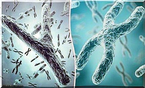
Autosomal Chromosomal Disorders
Autosomal chromosomal disorders are common. They can cause serious birth defects because chromosomal information is present in every cell in our body. Each arm of a chromosome is divided into 4 regions and in each region each band is numbered in relation to its distance from the centromere.
The short arm is called “p” and the long arm is called “q”. For example, 1 q 23 indicates: chromosome 1, long arm, second region, third band. Autosomal chromosomal disorders often lead to the following disabilities:
- Mental and intellectual deficiencies
- Presence of dimorphic features and deformities
- Delayed growth
Autosomal chromosomal disorders and their classification
There are two main types of chromosomal abnormalities: numerical disorders and structural disorders.
Autosomal chromosomal disorders: numerical disorders
The most common cause of numerical disorders in chromosomes is usually nondisjunction. This consists in a failure of the chromosome pairs or the chromatids to separate in the first or second meiotic division or during mitosis.
The consequence of this abnormality is that the parts of the chromosome pair do not separate properly. Therefore, in some germ cells there may either be too many chromosomes or part of a chromosome may be missing.
If a germ cell with an extra chromosome joins a normal germ cell during fertilization, the resulting zygote will contain three copies of that particular chromosome. This phenomenon is called trisomy.
When a germ cell that lacks a certain chromosome joins a normal germ cell, only one strand of the chromosome pair remains and a so-called monosomy is created.
Nondisjunction can also occur during mitosis, after meiosis phases I and II and the formation of the zygote. This results in the child being born with cells that are a mixture of trisomic and normal, or monosomic and normal.
These patients are called mosaics. In addition, this chromosomal disorder almost always affects chromosome X (the mosaics of other chromosomes are usually not viable).
Abnormalities in the structure of the chromosomes
The most common abnormalities in the structure of chromosomes are deletion, duplication, inversion, and translocation.
- Deletion or suppression. The loss of a chromosome fragment. As a result, the affected chromosome lacks all of the genetic information that is stored in the lost fragment.
- Duplication. The presence of an extra fragment of chromosomes. Sometimes a deleted fragment can join the very end of a homologous chromosome. This abnormality is much less harmful than the loss of a fragment of a chromosome.
- Inversion. This results in the fragmentation of a chromosome due to two tears. Then the adhesion to the same chromosome follows, but in the opposite way. Generally, this type of change in genes does not result in an abnormal phenotype. However, this can have consequences for the next generation if the inverted chromosome joins a normal chromosome.
- Translocation. The transfer of a part of a chromosome to another non-homologous chromosome. Sometimes these translocations are reciprocal.
The structural abnormalities usually occur during the meiosis of one of the gametes (maternal or paternal).
Autosomal chromosomal disorders
The most important chromosomal abnormalities are briefly explained below:
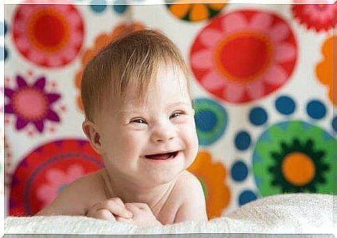
Down syndrom
It was first identified and described by Langdon Down in 1866. Down syndrome is the most common and well-known chromosomal disorder (it affects 1 in 700 births).
People with Down syndrome have 47 chromosomes and an extra chromosome 21 specifically.
This chromosomal disorder can usually be identified at or shortly after birth based on the following characteristics:
- Hypotension
- Mental disability
- Brachycephaly (overgrowth of the head due to premature closure of the coronal suture)
- Prominent tongue
- Short and thick hands.
- Heart abnormalities in 35% of cases
- The children are usually smaller than normal
Although 95% of Down syndrome cases are due to the trisomy of chromosome 21, about 4% have a translocation of the long arm of chromosome 21 (on chromosome 13, 14, 15, or 22). These patients have 46 chromosomes but are phenotypically indistinguishable from those with trisomy 21.
- The average age of mothers who have children with Down syndrome is 34 years.
- Male carriers are less likely to have children with the disease than women.

Trisomy 18 (Syndrome E)
This syndrome is associated with chromosome 18. It is associated with multiple congenital birth defects and is much more serious than Down syndrome (most patients do not live to be more than 6 months old).
The most common symptoms are:
- Mental disability
- Inability to grow
- Malformations of the ears, hands and feet
Trisomy 13 (Syndrome D)
This chromosomal disorder is less common than the previous syndrome, but the abnormalities are much worse (infants usually do not have a chance of survival beyond the first month of life).
Trisomy 13 causes nervous system defects, intellectual disability, cleft palate, cleft lip, skin abnormalities, heart defects, etc.
Cri-du-Chat Syndrome
This is due to a partial monosomy of chromosome 5 (the suppression of a fragment of the short arm).
Various anomalies occur here, the most characteristic of which is the infant’s cry. The child sounds like a meowing cat. Other deficiencies are intellectual retardation and microcephaly.
Monosomy 4
This is caused by the deletion of the short arm of chromosome 4. The disorder manifests itself in a phenotype characterized by low birth weight, a lack of growth, and an abnormal appearance on the face.
Autosomal Chromosomal Disorders: Conclusions
In summary, doctors struggle day in and day out to improve our knowledge of autosomal chromosomal disorders, which unfortunately affects many families and babies. Fortunately, they are always developing new ways to identify these conditions early on.
In addition, of course, the treatments for these abnormalities have evolved so there are now better ways to prevent them. At the same time, the care for children born with these complaints has also improved.